Services Performed
Colonoscopy
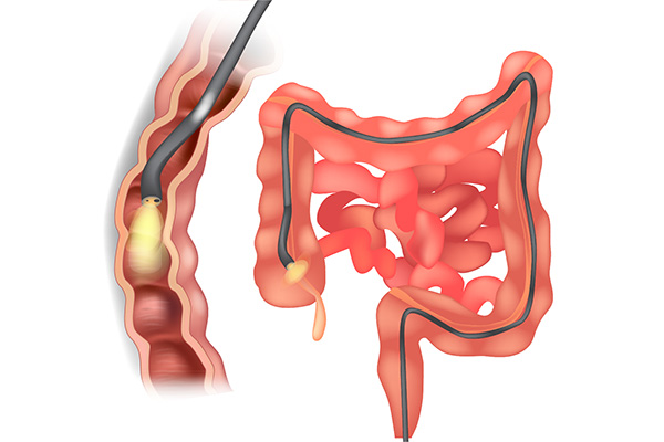
This procedure uses a colonoscope, a flexible tube-like device outfitted with a miniaturized camera, to take pictures of the colon. The colonoscope allows a physician to obtain a small tissue sample, apply medications or heat treatment directly to the lining of the colon, or remove polyps. Polyps are abnormal growths in the lining of the colon, which can become cancerous. Removing polyps is an important way to prevent colon cancer. A colonoscopy takes from 10 to 30 minutes. During the procedure, patients may feel some discomfort but rarely any pain. Patients receive medication intravenously to make them relaxed and drowsy. The span of service in the Endoscopy Center may last two to three hours, from admission through discharge.
Colonoscopy is a safe, simple and effective way to check for cancer, and to treat suspicious colon polyps. Screening is important because in the early stages of many colorectal diseases symptoms are not present. Cancer growth can go on for years and even decades within the colon, and is far more difficult to treat when symptoms do become present. The American Cancer Society recommends all men and women age 50 and older have a screening colonoscopy to check for signs of colon cancer, even if no symptoms are present. People with a family history of colon cancer should begin screening at an earlier age.
While the exact causes of colorectal cancer are not yet known, you are at a higher risk of Colorectal Cancer if:
- You are Over 50 years old
- You Eat a Diet high in animal fat
- You smoke or drink alcohol regularly
- You have a family history of colorectal cancer
- You are overweight
The Results
- Identify and Remove any potentially cancerous polyps within the lower intestines
- Detect inflammation, diseases and conditions of the colon
- Find the cause of bleeding or pain from the lower gastrointestinal tract
- Assess changes in bowel habits, such as chronic diarrhea
- Reassure your personal intestinal health or begin critical treatment
Upper Endoscopy
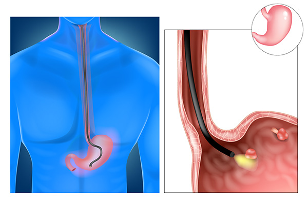
The Term “EGD” stands for esophagogastroduodenoscopy, also known as upper endoscopy, and is a procedure usually performed by a gastroenterologist (GI or intestinal doctor). This test involves passing a long flexible black tube with a light and video camera on one end through the mouth to examine and visualize the esophagus (the swallowing tube leading to stomach), stomach and the first part of the small intestines called the duodenum. The high quality picture from the endoscope is shown on a TV monitor and gives a detailed view of the upper GI tract. Specially trained nurses and technicians who are essential to performing the procedure safely and effectively will assist your doctor. The EGD takes about 5 to 10 minutes. The EGD can be helpful in the evaluation of many symptoms, such as: difficult or painful swallowing, abdominal pain, weight loss, heartburn, nausea, vomiting, anemia and the presence of blood in your vomit or stool.
Endoscopic treatment can be performed at the time of procedure. Examples of such treatments include dilation of the esophagus (stretching the esophagus), usually due to esophageal strictures, Schatzki’s Ring, Achalasia, Ingestion of caustic Agents and Tumors. Biopsies (taking small pieces of tissue) of any abnormality may also be done directly through the endoscope. Endoscopic biopsies are very superficial, taking only the very top layer of the esophagus, stomach or duodenum. Therefore, significant bleeding from the biopsies is rare and the biopsies do not scar the esophagus, stomach or duodenum.
You will be asked not to eat or drink anything for six to eight hours prior to your EGD. In the endoscopy unit, an IV (intravenous) line will be started in your arm or hand. You will be placed on a monitor that checks your heart rate, blood pressure and oxygen level, which is monitored by a certified nurse anesthetist (CRNA) who will be with you during the procedure. The medication that you will receive through your IV will make you very comfortable for the procedure. The sedation type is called conscious sedation, NOT general anesthesia. The main goal of this sedation is to make you comfortable, less anxious and diminish gagging. After sedation, the doctor will pass the endoscope through your mouth, most people spontaneously swallow and the scope is easily passed into the esophagus. Using the endoscope, the doctor can see a magnified picture of the lining of the upper intestinal tract on a video monitor. The EGD is a very safe and one of the most frequently performed procedures. It is the most sensitive test for detection of abnormalities of the upper GI tract lining.
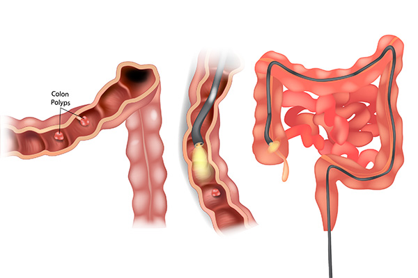
Sigmoidoscopy
A flexible sigmoidoscopy is an exam used to evaluate the lower part of the large intestine (colon).
During a flexible sigmoidoscopy procedure, a thin, flexible tube-like device outfitted with a miniaturized camera, is used to take pictures of the sigmoid colon. The physician is able to view the inside of the rectum and most of the sigmoid colon—about the last two feet (61 centimeters) of the large intestines. This procedure doesn’t allow the doctor to see the entire colon.